HarmonyCVI is a next-generation cardiac MRI analysis platform designed to deliver fast, accurate, and automated diagnostic insights. It streamlines cardiac imaging workflows using advanced computer vision and deep learning to support precise clinical decision-making.
AI & Deep Learning
Our AI powered Vision Analytics tool delivers state-of-the-art reliable performance in terms of Accurately generated Contours, Robustness & Computational Time
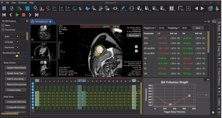
Volumetric Analysis
Volumetric assessment of Left & Right ventricles as well as Robust Segmentation & Visualization of LV & RV wall contours
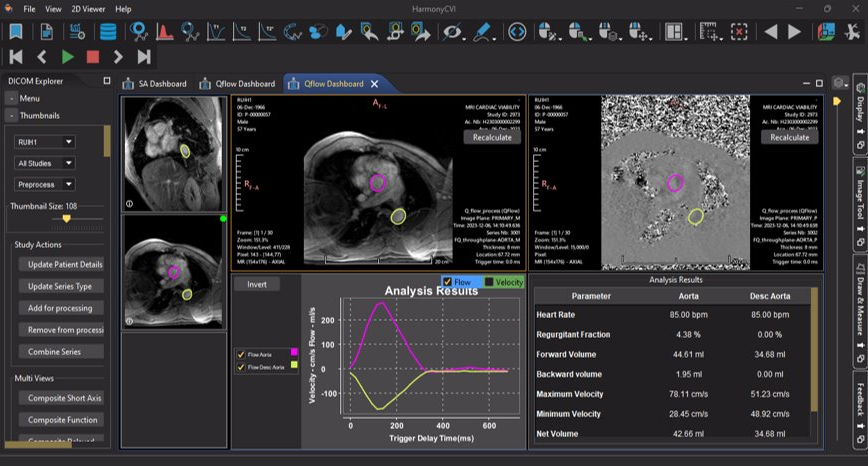
Q-Flow Analysis
Fully automated calculation of the volume & velocity of the blood flow without the need of contour drawing by user
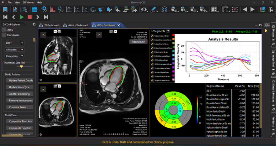
Strain Analysis
Accurate and reproducible myocardial strain and strain rate calculation and quantitative assessment of global, regional, & Myocardium function
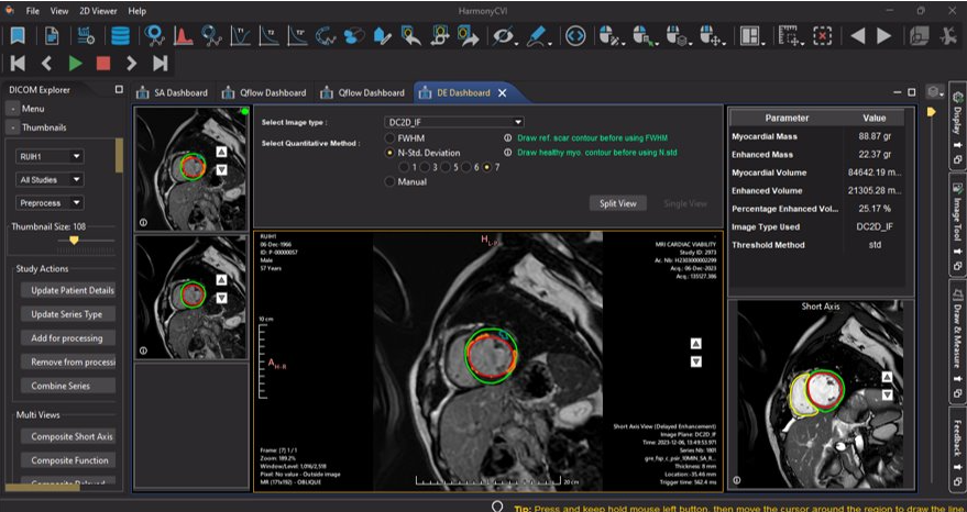
Fibrosis Quantification
Manual scar quantification process, incorporation of STD and FWHM for scar analysis as well as accurate analysis of scar percentage
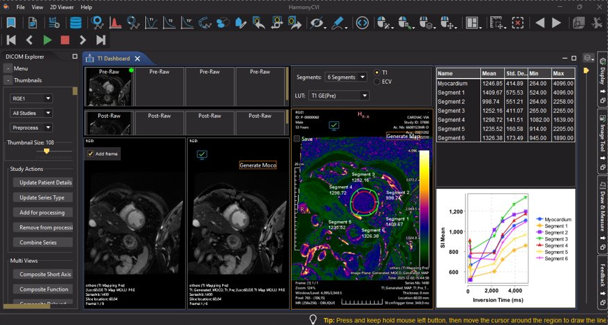
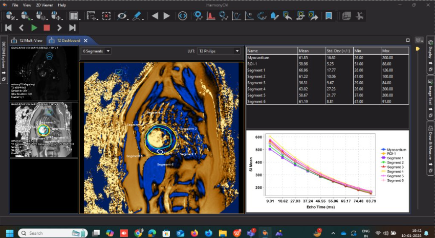
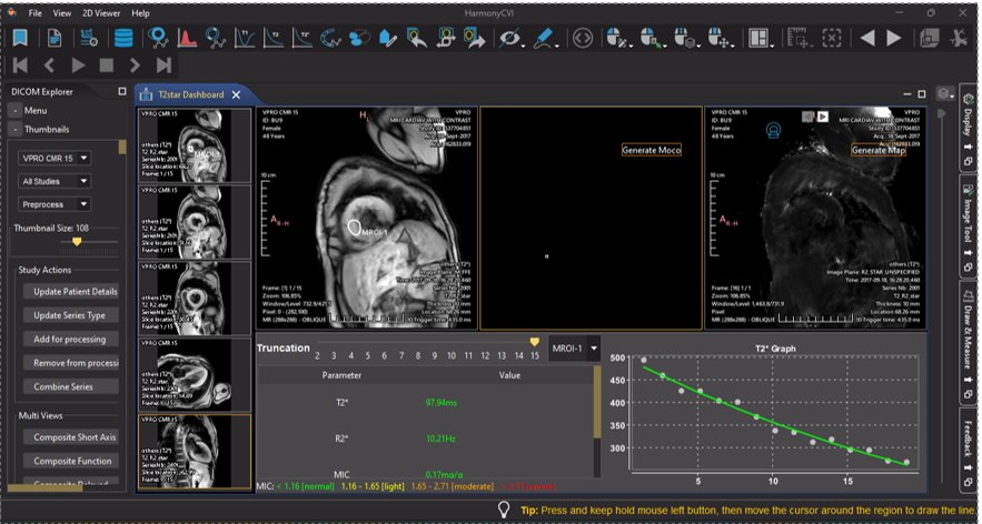
Myocardial Mapping
Precise quantification of T1, T2, T2*, and extracellular volume (ECV) helps detecting and assessing fibrosis, edema, iron deposition, and other cardiac tissue abnormalities with ease.
Cardiac MRI Analysis Capabilities
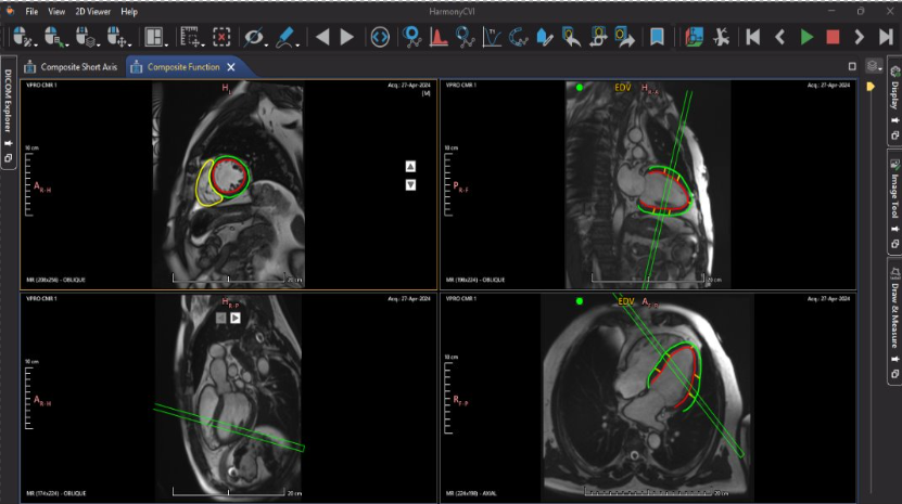
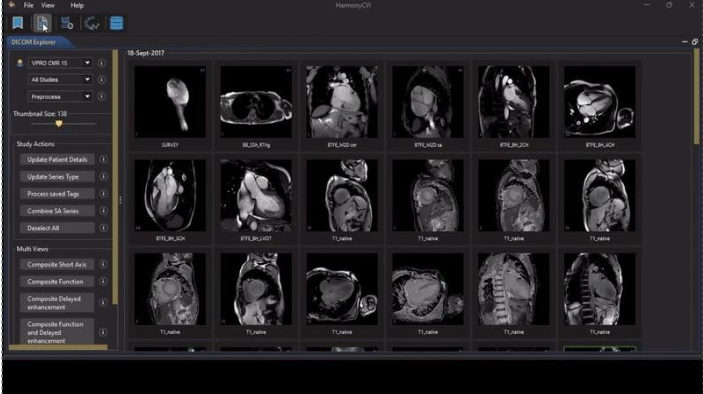
Multi-View Imaging & Synchronized Visualization
Automated combined view of critical cardiac MRI images with synchronized functional views, configurable layouts, and composite multi-panel displays to improve interpretation accuracy through simultaneous visualization.
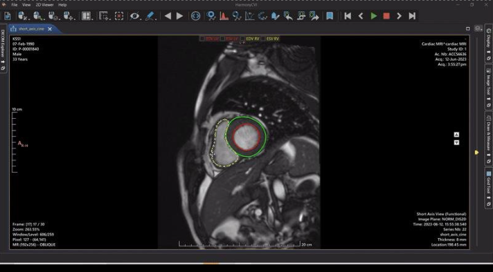
AI-Based Volumetric Analysis
Grid view with contour and EDV/ESV frame information, highly accurate deep learning–based contour detection, and comprehensive volumetric assessment of atria and ventricles with robust wall segmentation and continuous learning enhancement.
Automated Q-Flow Analysis
Automatic vessel segmentation with fully automated flow parameter calculations, graphical representation of flow and velocity, and quick, accurate output generation without manual intervention.
Delayed Enhancement & Scar Quantification
Contour generation using short-axis functional data with manual scar quantification options, incorporation of STD and FWHM methods, and precise scar percentage analysis for improved tissue characterization.
T1 Mapping & Extracellular Volume (ECV)
Flexible ROI definition, quantitative value measurement, graphical analysis, and automated ECV calculation for comprehensive myocardial tissue assessment.
T2 Mapping – Myocardial Edema Detection
Quantitative measurement of myocardial water content with absolute T2 relaxation values and color-coded parametric maps for objective edema and inflammation assessment.
T2* Mapping – Iron Overload Assessment
Precise quantification of myocardial and liver iron deposition using T2* relaxation times, supported by clear visual maps showing iron distribution.
Global Longitudinal Strain Analysis
Accurate and reproducible myocardial strain and strain rate calculations with automatic and manual LV boundary delineation, quantitative regional function assessment, and bull’s-eye visualization.
Secure Bookmarking & Workflow Continuity
Edit once and save securely, resume work across sessions, customize sharing with other users, and ensure protected access control for stored analyses.
Interactive Reporting & Clinical Documentation
One-touch upload of clinical data (PDF or text), real-time interactive report generation, structured and configurable reporting formats, customizable graphical representation, and integrated image inclusion for comprehensive diagnosis.